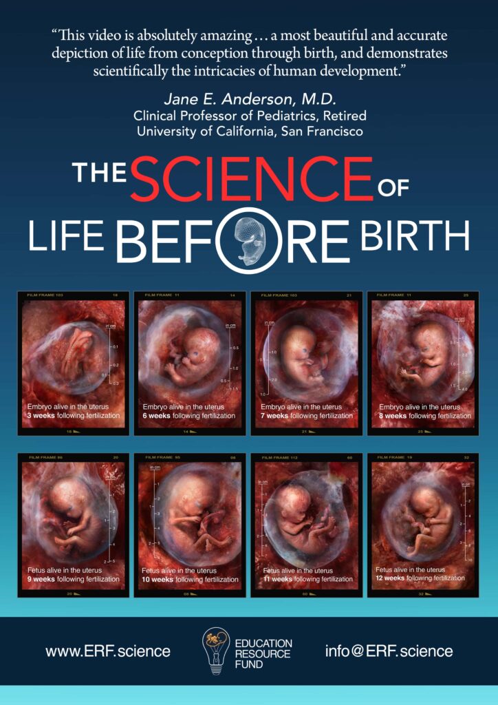
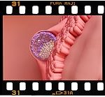
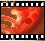
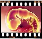
Endoscopic video of human embryos 3 weeks, 6 weeks, and 7 weeks following fertilization. The footage in this section contains no animation. It is actual prenatal medical imagery.

This image is available in 49 languages. Post on social media.


2023 Viddy Awards
Gold Winner

2023 Accolade Global Film Competition
Award of Excellence
Documentary Program Series
2023 Accolade Global Film Competition
Award of Excellence
Educational/Instructional/Training

2024 Creative Excellence Award
Website Design
An Education Resource Fund documentary titled The Science of Life Before Birth has been named a 2023 Viddy Gold film award winner. The Viddy Awards recognize “Outstanding achievement in video and digital skills.” The Viddy Awards competition is administered and judged by the Association of Marketing and Communication Professionals (AMCP) organization. This international group consists…

Awarded to ERF for our
Honoring Excellence In
Digital Creativity, Branding + Strategy
“AVA” stands for "audio-visual arts." The awards are presented through the Association of Marketing and Communication Professionals. The AMCP began in 1995 as a means to honor outstanding achievement and service in the communications profession.

Hailed as the “Internet’s highest honor” by The New York Times, The Webby Awards, presented by the International Academy of Digital Arts and Sciences (IADAS), is the leading international awards organization honoring excellence on the Internet. IADAS, which nominates and selects The Webby Award Winners, is comprised of Internet industry experts. The Education Resource Fund (ERF) has been honored for its See Baby Grow prenatal science app in the 2024 “Education, Science, & Reference” Webby Award category.
Physicians (especially OB/GYNs, pediatricians, and family practice doctors), as well as healthcare professionals generally, may wish to download the following Pregnancy Tracker video for display on a TV monitor in their waiting rooms and exam rooms, etc. This 4.5 minute, award-winning video features rare endoscopic and sonographic embryonic and fetal scans which span the entirety of pregnancy. The film can be programmed for looping on most TVs.
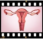
Please see below a comprehensive video archive containing nearly 22 hours of prenatal, endoscopic medical scans. Each scan depicts never-before-seen human embryos and fetuses, alive in the uterus, as they progress through each stage of prenatal development. The image bank features a searchable index listing approximately 6,000 of the anatomic structures and/or systems viewable in the video archive. Each search term is linked to the endoscopic video clip which images its corresponding structure and/or system, chronologically ordered by weeks following fertilization. The program is intended as a reference resource for use by clinicians, academics, researchers, and medical and nursing students, as well as students in the health sciences at undergraduate and graduate levels of study. The architecture of this interactive system is designed to accommodate continuous expansion.
Biologically speaking, “human development begins at fertilization,” when a woman and a man each combine 23 of their own chromosomes through the union of their reproductive cells. The DNA in the 46 chromosomes of the resulting embryo (zygote stage), then only one cell in size, already contains some 3 billion base pairs of digital data, the genetic blueprint for the entire human body...

The human heart will beat 3 billion times over the course of an average lifespan.
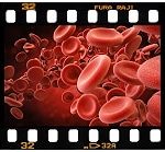
The human circulatory system contains 20-30 trillion blood cells at any given time.
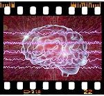
The human brain contains 100 billion neurons.
The neurons in the human brain are linked to one another by 100 trillion synaptic connections.
High-resolution images of embryos and fetuses developing in utero


")


Follow the Science - Fertilization through 12 weeks


“By convention, obstetricians date pregnancy from presumed first day of the last normal menstrual period (LMP). This is gestational age, which in embryology is superfluous because gestation does not begin until fertilization of an oocyte occurs. Embryonic [or fetal] age [also described as fertilization or conceptional age] begins at fertilization, approximately 2 weeks after the LNMP…. The day on which fertilization occurs is the most accurate reference point for estimating [embryonic or fetal] age ….” The Developing Human, Clinically Oriented Embryology, Moore, Persaud & Torchia, Elsevier, 10th Ed. (2016). Unless otherwise noted, all embryonic and fetal ages in Education Resource Fund curricular materials are estimated in weeks/months following fertilization.

Science Documentary Films


The Science of Life Before Birth
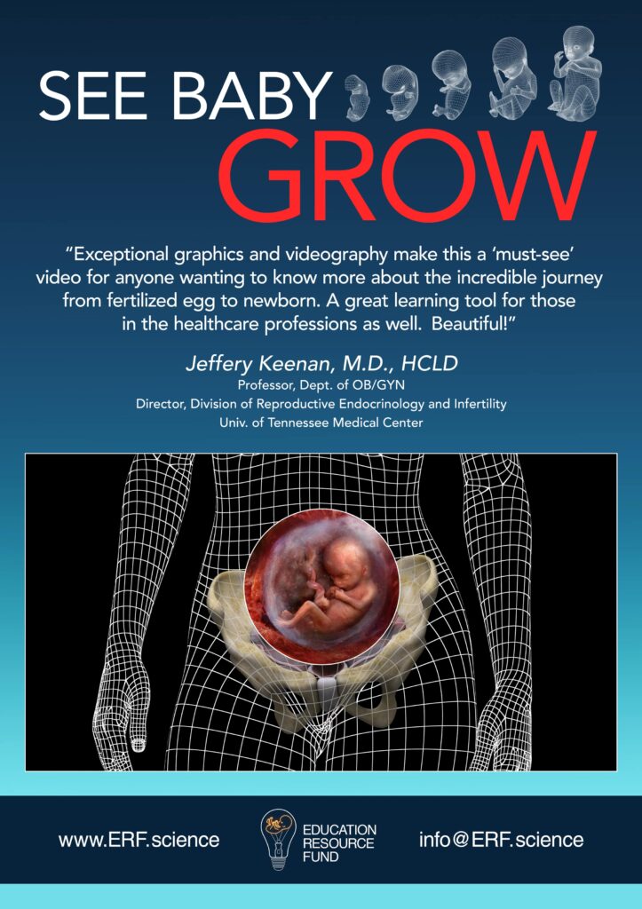
See Baby Grow
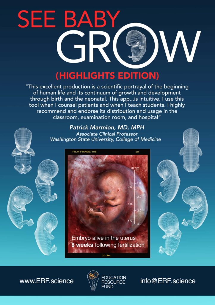
See Baby Grow (Highlights Edition)
The Science of Life Before Birth – Spanish
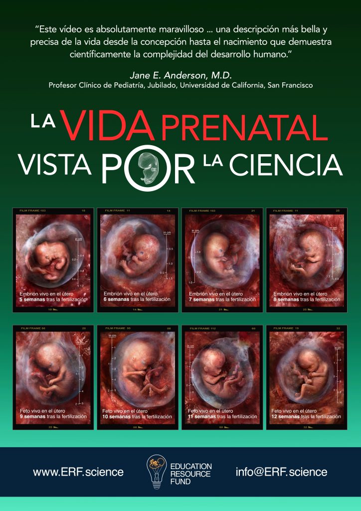
See Baby Grow – Spanish
See Baby Grow (Highlights Edition) – Spanish
AWARD-WINNING PREGNANCY TRACKER

The Intricately Interactive Choreography of Conception
1. Male initiated:
Up to 600 million sperm are deposited in the birth canal, of which only 200 reach the fertilization site in the uterine tube. The Developing Human: Clinically Oriented Embryology, 10th ed., K. Moore et al., Elsevier (2016), pp. 25-26.
2. Male initiated: An enzyme produced by...
ERF’s See Baby Grow app content has been viewed more than 13.6 million times as of April 5, 2024. More than 1,400 comments have been posted. Viewers are from at least 173 countries.

© See Baby Grow App
To obtain the See Baby Grow App for Apple (iOS), download from the Apple App Store at the foregoing QR Code, or this link: https://apps.apple.com/us/app/see-baby-grow/id1633494975.
To obtain the See Baby Grow App for Google Play (Android), download from Google Play App Store at the foregoing QR Code, or this link: https://play.google.com/store/apps/details?id=com.seebabygrow.erf.

Public Service Announcements


Make Science Fun for Kids

Children’s Science Documentary Films


Before You Were Born
Before You Were Born – Spanish
Before You Were Born – Chinese (Simplified)
Before You Were Born – Chinese (Traditional)
Before You Were Born – Hindi
Before You Were Born – English (Indian Narrators)
Coloring Books
Color using your finger on a phone, or your finger or stylus on a tablet, or your mouse and cursor on a computer, or your crayons on physical pages you print out on paper.


Auto-fill zone coloring
Color with finger or stylus
"I hold a multiple subject teaching credential in the state of California and I have been a public elementary school teacher since 2004. My primary focus has been Kindergarten, First and Second grade.
I recently examined the ERF coloring pages which depict embryos and fetuses developing in utero. This is an amazing interactive resource for children of all ages. It's extremely user friendly. The high-resolution prenatal images next to the coloring book line drawings are fantastic! This is an instructive resource that can be used effectively in the classroom setting.
I also reviewed the ERF site where I watched the children's version of the ERF prenatal video. Like the coloring pages, the children’s edition of the ERF prenatal science documentary is also an amazing resource! All content is age-appropriate for even the youngest children."
Ellarose Pinkus
Puzzles


WORD SEARCHES


Curricular Content


Embryoscopy, fetoscopy, and high-resolution ultrasound imagery, showing embryonic and fetal development.
 Pregnancy Week 1
 Pregnancy Week 2
 Pregnancy Week 3
 Pregnancy Week 4
 Pregnancy Week 5
 Pregnancy Week 6
 Pregnancy Week 7
 Pregnancy Week 8
 Pregnancy Week 9
 Pregnancy Week 10
 Pregnancy Week 11
 Pregnancy Week 12

 Pregnancy Month 4
 Pregnancy Month 5
 Pregnancy Month 6
 Pregnancy Month 7
 Pregnancy Month 8
 Pregnancy Month 9
Subtitles in 92 languages for ERF video
"The Science of Life Before Birth"
Choose Your Preferred Subtitle Language Here:
We have prepared the script of “The Science of Life Before Birth” in 92 different languages. You can download a PDF of each of those translations by following the links below.


See Baby Grow app video
The See Baby Grow app video depicts embryos and fetuses, alive in the uterus, throughout every stage of pregnancy. These preborn babies have been scanned using embryoscopy and fetoscopy medical imaging technology, as well as high-resolution, research-grade sonography. The narration describes developmental anatomy and physiology as it unfolds through all three trimesters of pregnancy.
If you are a medical practitioner or science educator/researcher, or a student, please consider offering an endorsement for this project.
PROFESSIONAL ENDORSEMENTS
Professional reviews neither state nor imply institutional endorsement.
Post the following prenatal development facts on your social media:


The ERF prenatal videos featured at www.ERF.science are updated and expanded variants of the science documentary titled "The Biology of Prenatal Development."
Biology of Prenatal Development Film Awards

CINE Golden Eagle

Platinum Remi Award

Grand Remi - "Best of Show"

Best of Show | Award of Excellence | Award of Excellence

Grand Remi

Award of Excellence

Silver Telly Award
Principal Technical Advisor:
Mark T. Cullen, M.D.
Technical Review:
James H. Baker, Ph.D.
Enid Gilbert-Barness, M.D.
David H. Bernanke, Ph.D.
Mark J. Holterman, M.D., Ph.D.
David L. Bolender, Ph.D.
Paul A. Krieg, Ph.D.
Professor Stuart Campbell, D.Sc.
Maria Michejda, M.D,
Bruce M. Carlson, M.D., Ph.D.
Maurice J. Pescitelli Jr., Ph.D.
Julian E. De Lia, M.D., M.B.A.
Charles L. Saxe, Ill, Ph.D.
Charles H. Ellis Jr., Ph.D.
Mark F. Seifert, Ph.D.
Ona Marie Faye-Petersen, M.D.
Allan R. Sinning, Ph.D.
David W. Fontaine, M.D.
Bradley R. Smith, Ph.D.
Ravmond F. Gasser, Ph.D.
Sam R. Voora, M.D.
ENDORSEMENTS OF THE BIOLOGY OF PRENATAL DEVELOPMENT
“Truly unlike anything I’ve seen before in the area of human development. The graphics, mixed with tape of actual intrauterine developing fetuses, makes the biology of human development…..human! I would offer it to both science…
Julian E. De Lia, M.D., M.B.A.
Medical Director
The International Institute for the Treatment of Twin to Twin Transfusion Syndrome
St. Joseph Regional Medical Center*
Clinical Associate Professor, Department of Obstetrics & Gynaecology
Medical College of Wisconsin*
“This video provides a splendid review of prenatal human development from fertilization to birth. The intrauterine videophotography and imaging is exquisite and breathtaking and allows the viewer to witness the different stages and external features…
Mark F. Seifert, Ph.D.
Professor of Anatomy and Cell Biology
Indiana University School of Medicine*
“This is a well conceived and executed video. The use of different types of images is well thought out and effective. The text is accurate and well chosen. This video is particularly appropriate for an…
Charles L. Saxe, III, Ph.D.
Associate Professor
Department of Cell Biology
Emory University School of Medicine*
“The Biology of Prenatal Development does a wonderful job of presenting and describing the milestones of human development from conception to birth. The video sequences and animations provide beautiful visual representations of the concepts described through…
Bradley R. Smith, Ph.D.
Associate Professor and Director, Biomedical Visualization Graduate Program
School of Art and Design
University of Michigan*
Senior Associate Research Scientist
Department of Radiology
University of Michigan Medical School*
“This video is a significant contribution for the education of the general public about human prenatal life. The many colored images of living, moving embryos and fetuses reveal the remarkable beauty of life inside the…
Raymond F. Gasser, Ph.D.
Emeritus Professor, Cell Biology and Anatomy
Adjunct Professor, Obstetrics and Gynecology
Louisiana State University Health Sciences Center*
“An excellent program. Should be very useful for pathologists, obstetricians, ultrasonographers, [and] perinatologists.”
Enid Gilbert-Barness, M.D
Professor of Pathology & Laboratory Medicine
Professor of Pediatrics
Professor of Obstetrics & Gynecology
University of South Florida College of Medicine*
“The Biology of Prenatal Development video provides a uniquely accessible visual overview of prenatal human development.”
Paul A. Krieg, Ph.D.
Professor of Cell Biology & Anatomy
Professor of Molecular & Cellular Biology
The University of Arizona College of Medicine*
“After reviewing this video, I find it to be a first class presentation that would ideally set the stage for the study of development at all levels of education.”
Alan R. Sinning, Ph.D.
Associate Professor of Anatomy
University of Mississippi Medical Center*
“This beautifully produced video provides an impressive overview of human development from conception to birth in a very clear, concise presentation. The informative and engaging format includes a continuous timeline to emphasize the sequence and…
David H. Bernanke, Ph.D.
Associate Professor of Anatomy
Medical University of South Carolina*
“This visually compelling video provides important insights into the dynamism of the developing human embryo. The images can be used to supplement existing teaching tools in courses on developmental biology.”
Mark J. Holterman, M.D., Ph.D., FAAP, FACS
Assistant Professor of Surgery
Division Chief, Pediatric Surgery
University of Illinois College of Medicine at Chicago*
“The Biology of Prenatal Development is an excellent overview of key features of human embryonic development. It contains amazing video sequences of human embryos combined with helpful animations and very understandable narration. It reveals the mystery…
David L. Bolender, Ph.D.
Associate Professor, Department of Cell Biology, Neurobiology and Anatomy
Graduate Faculty, Program in Cell & Developmental Biology
Medical College of Wisconsin*
“This video production is a uniquely crafted portrayal of human embryofetal development that is completed by a succinct, but very accurate, description of the process. It is a joint effort that has utilized the expertise…
Ona Marie Faye-Petersen, M.D.
Associate Professor of Pathology and Obstetrics & Gynecology
University of Alabama School at Birmingham*
Head, UAB Microdissection Laboratory for Perinatal Pathology
Department of Pathology, Anatomic Division
“The Biology of Prenatal Development provides an amazing view of human embryonic and fetal development. It would be a great supplement to developmental biology courses at the undergraduate, graduate, and medical school level.”
Deborah J. Andrew, Ph.D.
Associate Professor
Department of Cell Biology
Johns Hopkins University School of Medicine*
“The combination of exquisite images and a clear, carefully researched text makes this video an unparalleled resource for all who are interested in the mysteries of life before birth.”
Bruce M. Carlson, M.D., Ph.D.
Director and Research Professor, Institute of Gerontology
University of Michigan*
Professor, Cell and Developmental Biology
University of Michigan Medical School*
“The developmental period before birth is increasingly understood as a time of preparation during which the developing human acquires the many structures, and practices the many skills, needed for survival after birth. As our understanding of early human development advances, so too will our ability to enhance health––both before and after birth.”
The Biology of Prenatal Development, a documentary film originally distributed by the National Geographic Society
The following science documentaries, medical textbooks, and medical journal articles are among the many educational resources which provide useful information regarding the biology of prenatal development:
Recommended Science Documentaries


Recommended Medical Textbooks


Medical Journal Articles
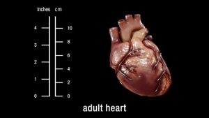
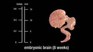
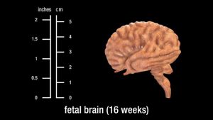
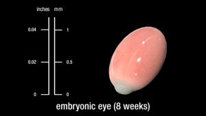
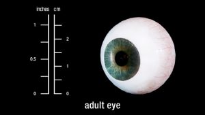
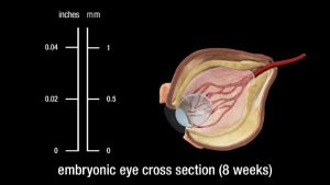
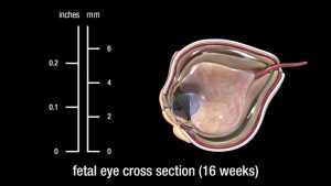
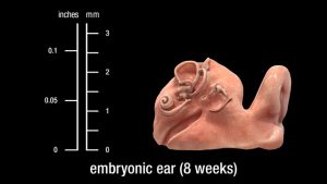
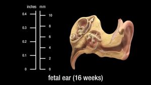
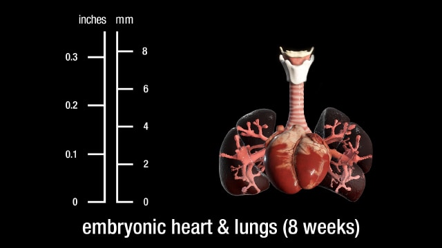
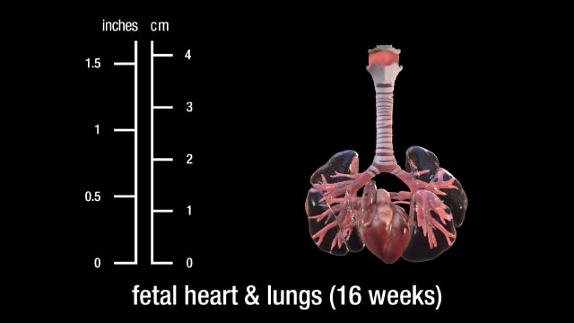
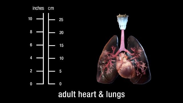
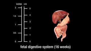
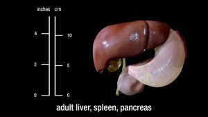
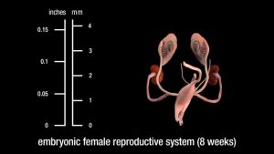
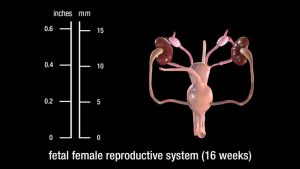
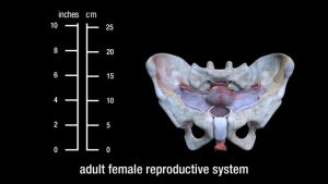
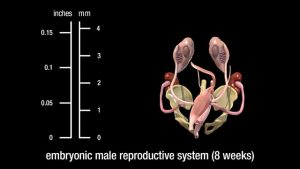
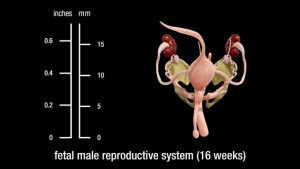
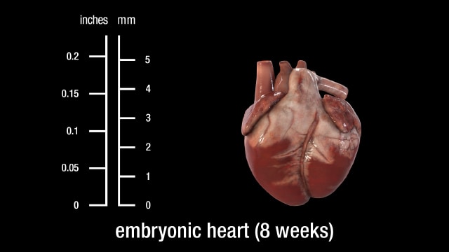
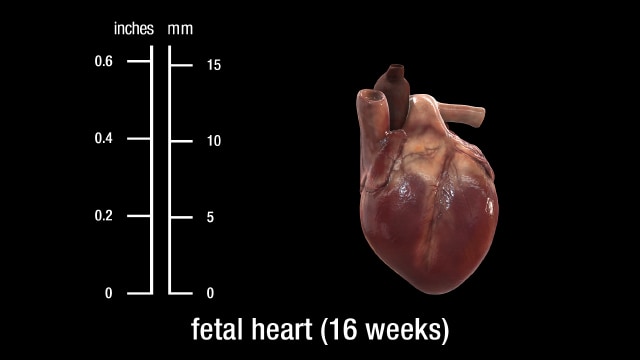
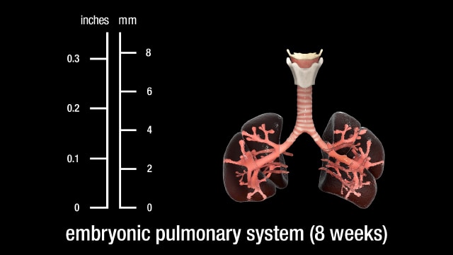
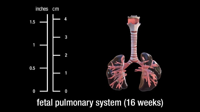
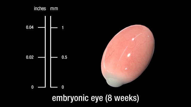
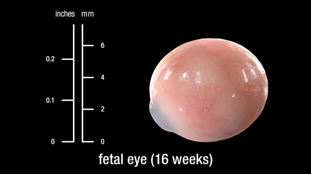
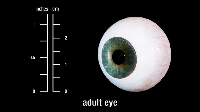
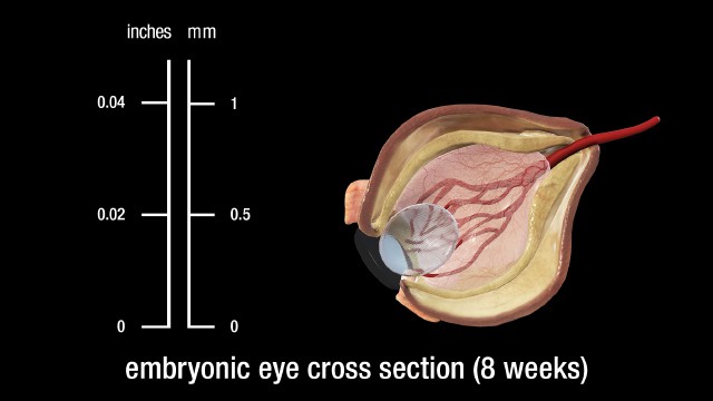
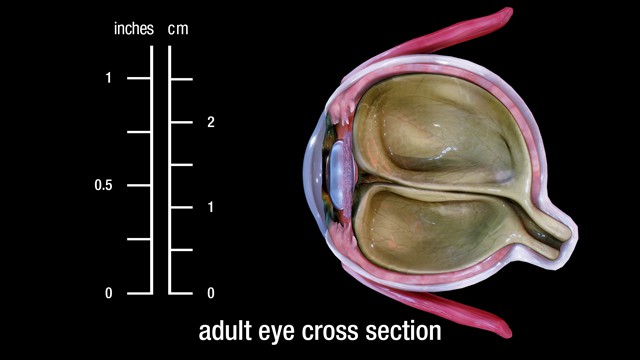
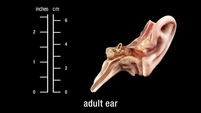
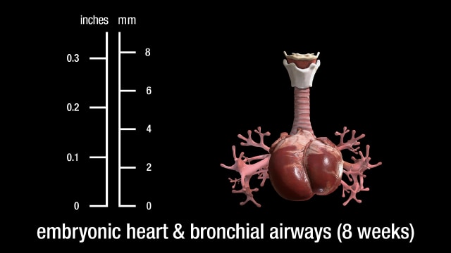
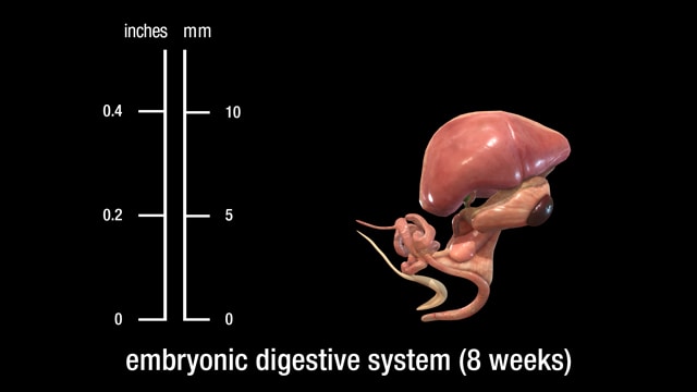
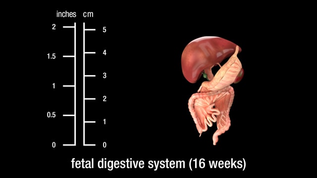
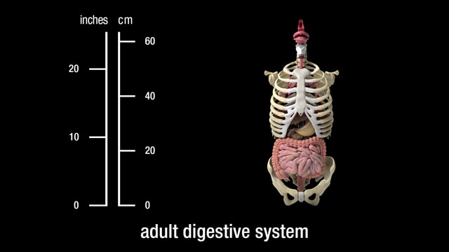
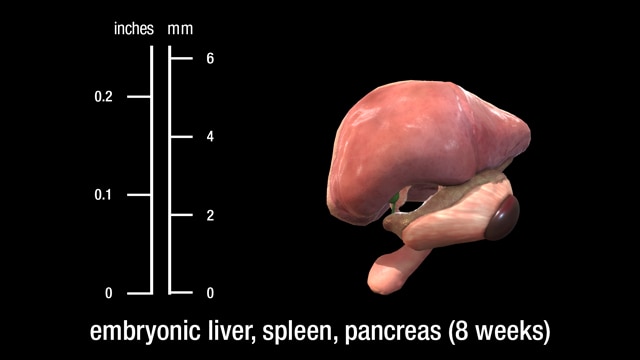
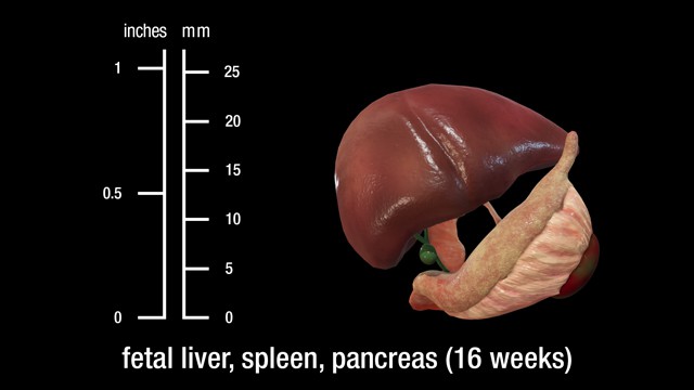
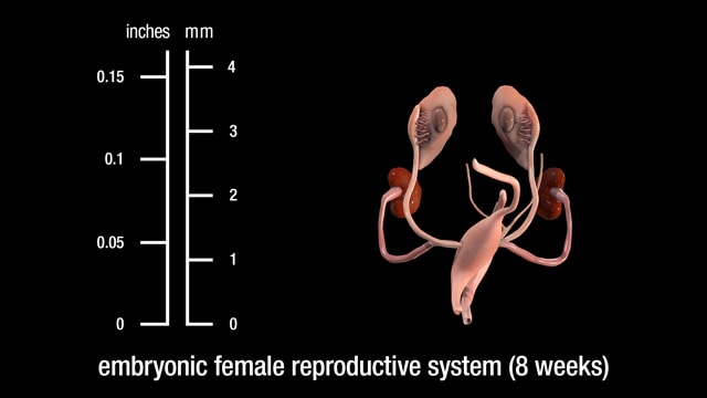
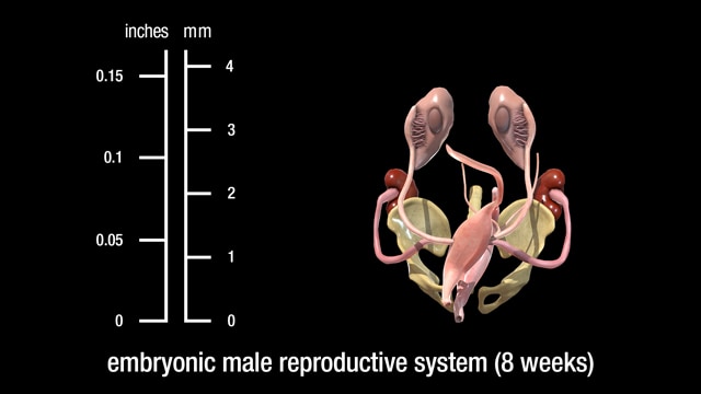
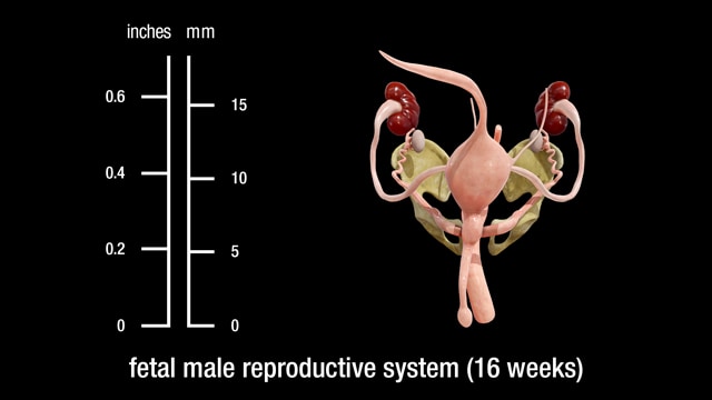
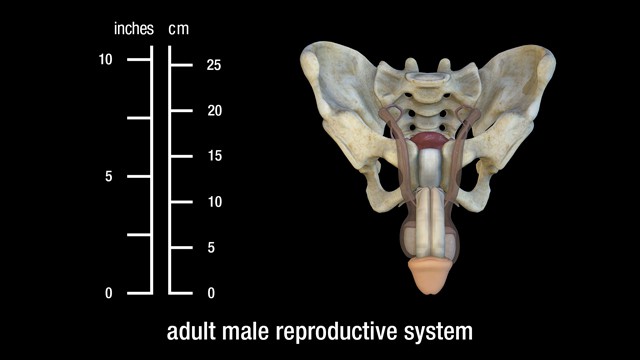
Comparative Embryonic and Fetal Anatomy
See 12 of your baby's vital organs depicted in embryonic, fetal, and adult age stages and illustrated as 3D models which you can rotate about their vertical, horizontal, etc. axes


IMAGING THE UNIMAGINABLE
The Education Resource Fund’s (ERF) composited embryonic and fetal pictures are derived from many smaller images, “stitched” together in much the same manner employed by NASA to combine satellite photo “tiles,” in a process which forms a large “mosaiced” image. Many research institutions use related technology to image otherwise unimageable (and unimaginable) objects and processes.

A mosaic image of the lunar South Pole, with individual photo tiles electronically stitched together to create one enormous panoramic view. Photo credit NASA
ENDOSCOPES
ERF’s embryo and fetus imagery was initially derived by teams of physician researchers and clinicians employing endoscopy (and its subsets, embryoscopy and fetoscopy) to diagnose and treat prenatal disorders in utero.

An endoscope with related equipment.
Endoscopes are medical imaging devices which permit high-resolution observation of tissues and processes inside the human body. Prenatally, they can be used to produce minimally invasive scans imaged through, but from outside, the amnion. When clinically necessary, more invasive scans may be performed by surgically entering the abdominal cavity, uterus and amniotic sac. At the distal end of these instruments is an objective lens designed for imaging. At the proximal end is an eyepiece, or sensor, which enables viewing.
HOW THEY WORK
These scopes generally consist of a tube which encloses a relay lens system (in rigid endoscopes) or a fiber bundle (for fiber-optic, or flexible, endoscopes) for illumination and to transmit an image from the objective lens inside the body to the proximal end outside.
Said differently, endoscopes use optical elements to direct light to the area sought to be illuminated and transmit the resulting image to the eye or detector. Rigid endoscopes generally offer superior resolution or magnification. But an endoscope’s objective lens is only approximately 1/5 of an inch in diameter, and this relatively small size substantially narrows the observer’s field of view (even with the addition of supplemental lenses such as “negative” or “prism” optics, etc.).
CONSTRAINTS
This limitation is further compounded by the need to use the scope in very confined spaces, with only short distances separating the objective lens from the anatomical structures being imaged. Consequently, only a small segment of the embryo or fetus is observable at any point along the timeline of the scan.
An endoscope’s construction must also accommodate frequently conflicting design considerations. The resulting compromises can involve not only fields of view, but depths of field (meaning thickness of the plane of focus) and image illumination and magnification, as well as distortion issues (i.e., stretched or compressed perspective), etc.
WORK-AROUNDS
Therefore, to produce a high-quality, single image of the entire embryo or fetus, large numbers of smaller, more detailed pictures must be joined together in a manner suggestive of the process by which puzzle pieces are assembled to form a completed picture.
This technique employs a complex proprietary process which combines segmental scans to create a final, multi-source composited image. The resulting picture is digitally adjusted to preserve each segment’s original color, resolution, contrast, proportions, illumination, etc. Technicians also correct for vignetting (image degradation involving content loss at the periphery of the frame).
MAGNETIC RESONANCE IMAGING & ULTRASOUND
The British medical journal Lancet has published a prenatal magnetic resonance imaging (MRI) study similarly involving the creation of 3D pictures to diagnose and treat congenital heart problems in utero. The BBC reports that “A series of 2D pictures of the heart are taken from different angles using an MRI machine” to image the fetus.

Magnetic Resonance Imaging (MRI) equipment with digital images of scanned tissue.
The story explains that “Sophisticated computer software pieces the images together, adjusts for the beating of the heart and builds … [a] 3D image of the heart.” A pediatric cardiologist describes the resulting 3D images as “beautiful.”
This MRI research is part of a fetal diagnostic project which is also exploring scans using “four ultrasound probes at the same time – current scans use one – to get a more detailed picture.” This process produces a more wholistic composited image.

Research-grade ultrasound scanning equipment.
NASA COMPOSITES IMAGERY (SINGLE MEDIUM)
ERF’s imaging process is conceptually similar to the technologies used by the National Atmospheric and Space Administration (NASA) to produce wide-area satellite images of the earth’s surface. Until the launch of the Deep Space Climate Observatory Satellite (DSOVR), which now orbits the earth at a distance of one million miles, NASA had no camera positioned sufficiently far from our planet to capture the globe’s entire sunlit surface in a single photograph.
As previously noted, an endoscope’s objective lens must also operate too near to an embryo or fetus to permit its entire anatomy to be imaged in a single frame. This same constraint complicates the capture of satellite imagery. Previous pictures of the earth could, therefore, only be created using digital “stitching” technology to make one large composite image from many smaller segments. Scientists sometimes describe this final image (or “data set”) as a “mosaic,” comprised of large numbers of individual tiles.
HYBRID IMAGERY (MULTI-MEDIA)
A satellite picture can also be augmented by aerial photography (cameras on aircraft operating within the earth’s atmosphere) to improve resolution. Hybrid images of this sort can be created by superimposing black and white imagery (for still higher resolution) over color pictures of the same area, the latter to optimize chromic (color) fidelity.
In this connection, the scientific press reports that the Landsat Image Mosaic of Antarctica (LIMA) “combined over one thousand precise, calibrated satellite images with other data from the continent’s surface to create a single picture of the entire continent.” The high magnification factor (think telephoto lenses which enlarge image objects) of each of these puzzle pieces yielded a composite picture depicting more detail than could have been captured in a single photo shot with a wide-angle lens.

Landsat satellite cameras generate composited images of the earth’s surface during multiple passes with continuous scans per pass.
APPLICATIONS IN ASTRONOMY
NASA uses this mosaicking process to image celestial bodies of nearly every description. The Juno spacecraft made composite images of Jupiter; InSight of Mars; Cassini of Saturn; and Hubble of the Sombrero Galaxy.
An exquisitely detailed depiction of a challenging subject, whether prohibitively small or large, near or far, may involve vastly more complexity than meets the eye, and there exist nearly countless examples of comparably creative combinations of imaging techniques.
ENTAMOLOGICAL USES
NASA is not alone in its use of this imaging technique. A Wall Street Journal story, July 30, 2022, titled "Breathtaking Bugs at the Museum,” describes an exhibition of “macrophotography prints [which] makes us appreciate these small creatures’ beauty on a large scale.” The article explains the creation of large format prints “in an exhibit called ‘Extinct and Endangered, Insects in Peril.’” The imaging process is called “macrophotography,” and it took an average of “three weeks and as many as 10,000 files for the photographer to make an individual image.”

A digital camera with bellows, reversal rings, macro couplers, and focusing mechanism, typically used for extreme closeups of exceedingly small objects. Photo credit B&H Photo

A digital camera’s closeup macro rings.

A tube lens of the type used for extreme closeup photos of exceedingly small objects. Photo credit B&H Photo
The photographer used a digital camera “to which he attached a bellows, a tube lens and a microscope objective (basically a very high-powered magnifying glass).” The process requires that the “subject insect … be divided into as many as 20 or 30 sections, each of which is photographed separately.” Lighting is critically important “because eyes, legs, wings, etc. reflect light differently ….” The photographer consequently “devised different lighting regimes for each.”
Even more remarkably:
The macrophotography camera setup has an extremely shallow depth of field, so the lens is electronically advanced toward the subject in seven-micron increments (a micron equals one millionth of a meter) taking 400 or 500 digital pictures that are then merged to create an image in sharp focus from front to back.
The article adds that, “Paradoxically, smaller insects require more files than larger ones.” In the aggregate, “the section images are joined to make a file that may be as large as eight, nine or 10 gigabytes … The detail is stunning ….”

A research-grade microscope.
A topically related story was posted by the journal Nature, March 10, 2023, titled, “Gigantic map of fly brain is a first for a complex animal.” First published in the journal Science, the project was made possible when:
Researchers spent a year and a half capturing images of the brain of a single six-hour-old Drosophila larva with a nanometre-resolution electron microscope. Using a computer-assisted programme, they then pinpointed the neurons and synapses and spent months manually checking them.

A high-power, electron microscope.
Science reported a related July 19, 2018, story titled, “In a ‘tour de force,’ researchers image an entire fly brain in minute detail.” The imaging methodology was, so to speak, mind-boggling:
[Researchers] soaked a fly’s brain in a solution containing heavy metals, which bind to the membranes of neurons and to proteins at the synapses …. [Next the team used] a diamond knife [to] cut the brain into about 7000 slices, each of which was struck with a beam of electrons from the microscope to create an image.
The process required a camera that could capture 100 frames per second, a robotic system to scoot each brain slice into place with nanometer precision, and software to stitch together the resulting 21 million pictures. The result is a reconstruction that lets researchers zoom in on the features of an individual synapse.
FROM THE SMALLEST TO THE LARGEST
The Orange County Register carried a similar article, February 18-19, 2023, titled, “See how the world’s largest photograph was created in Irvine [CA].” The picture involved 6 photographers working for 8 months to create a “panoramic view of … the former Marine Corps Air Station El Toro ….”

A commercial, pinhole camera, sometimes used for stitching together individual photos to create a pantographic image of one very large object. Photo credit marekuliasz/Shutterstock
Using an ancient “pinhole camera” concept, the process captured “images of every single building” on the 4,800-acre base. The picture was created from about 500,000 individual images, shot from distances of 50-75 yards between the camera and the object being photographed. Each negative was then painstakingly exposed on one solid piece of muslin cloth, 31 feet tall and 111 feet long. The end-result was a single, giant, composite, historically significant photograph of virtually the entire military facility.
IMAGING IMPLICATIONS FOR NEUROLOGY IN HUMANS
The journal EHP, Environmental Health Perspectives, November 20, 2018, published an article titled, “The Brain before Birth: Using fMRI [functional magnetic resonance imaging] to Explore the Secrets of Fetal Neurodevelopment.” In this case, the imaging technology is used to study the trillions of neural connections, called the connectome, which link the “billions of threadlike fibers [that] crisscross the brain.”
The author explains that “fMRI is not perfect,” and that the images “generated by the technology must be manipulated to correct for distortion and to scale brain scans to a consistent, comparable template.” Equally problematic is the fact that “technical issues potentially result in artifacts that may not be recognized as errors.”
OCEANOGRAPHIC RELEVANCE
Arstechnica.com/science, May 17, 2023, posted an essay titled “3D ‘digital twin’ showcases wreck of Titanic in unprecedented detail.” Previous film of the wreck employed low-resolution cameras, and a 1997 documentary produced by James Cameron used “miniature models and special effects … since Cameron couldn’t get the high-quality footage he needed for a feature film.” The new pictures of the ship were obtained using two submersibles as camera platforms, mapping
… every millimeter of the wreck, including the debris field spanning some three miles. The result was a whopping 16 terabytes of data, along with over 715 still images and 4K video footage. That raw data was then processed to create the 3D digital twin. The resolution is so good, one can make out part of the serial number on one of the propellers.
‘This model is the first one based on a pure data cloud, that stitches all that imagery together with data points created by a digital scan, and with the help from a little artificial intelligence, we are seeing the first unbiased view of the wreck.’
One of the producers explained to BBC News that “you have to map every square centimeter—even uninteresting parts, like on the debris field you have to map mud, but you need this to fill in between all these interesting objects.”

Submersible research craft with digital imaging equipment, generally used for deep sea exploration and composite photography. Photo credit maliao/Shutterstock
On the same topic, a May 17, 2023 insider.com story, headlined “First-ever full 3D scan of the Titanic on the sea bed reveals the ruined ocean liner in incredible detail,” describes the production as “the largest underwater scanning project in history.” It explains that “Previous footage has only allowed you to see one small area of the wreck at a time,” but this “model will allow people to zoom out and look at the entire thing for the first time ….”
METEOROLOGICALLY RELATED IMAGE PROCESSING
The Wall Street Journal, February 17, 2023, ran a story headlined “El Niño Likely to Form By Summer ….” It described the process by which weather forecasts are also made using composite imagery:
NOAA meteorologists make predictions of the ENSO cycle using statistical models that compare historical records with current ocean and atmospheric conditions. They also use computer models that combine data from satellites, ocean buoys, ships’ weather balloons and land-based stations into algorithms that form a picture, or map of what the future state of the atmosphere looks like based on the model’s calculations.

A weather satellite of the type currently used to collect meteorological data.
IMAGE PROCESSING MAKES INVISIBLE DEEP SPACE PHENOMENA VISIBLE
According to gizmodo.com, on July 12, 2022, “the first full-color images from the Webb Space Telescope showed countless nebulae, galaxies, and a gassy exoplanet as they had never been seen before.” The article, headlined “Are the Colors in Webb Telescope Images ‘Fake’?” answers an emphatic “NO!” As it turns out, “Webb only collects infrared and near-infrared light, which the human eye cannot see ….”
The story goes on to explain that:
Image developers on the Webb team were tasked with turning the telescope’s infrared image data into some of the most vivid views of the cosmos we’ve ever had. They assign various infrared wavelengths to colors on the visible spectrum, familiar reds, blues, yellows, etc. But while the processed images from the Webb team aren’t literally what the telescope saw, they’re hardly inaccurate.

James Webb satellite telescope used for extensively processed, deep space imaging.
By way of further explanation, “Astronomy is often done outside the visible spectrum, because many of the most interesting objects in space are shining brightly in ultraviolet, x-rays, and even radio waves.” Instruments such as Webb were “designed to extend the power of our vision, to go beyond what our eyes are capable of doing, to see light that our eyes are not sensitive to.”
Using infrared light to image objects enables astronomers to “penetrate thick clouds of gas and dust in space, allowing researchers to see previously hidden secrets of the universe,” and then convert that monochromatic light into wavelengths visible to the human eye. Even more image processing is then necessary because “Webb’s raw images are so laden with data that they need to be scaled down before they can be translated into visible light.”
The Wall Street Journal, July 13, 2023, published a spectacular image of the “Rho Ophiuchi cloud complex, the closest star-forming region to earth … seen in a composite of separate exposures acquired by the James Webb Space Telescope using its NIRCam instrument.” As described above, the original image was monochromatic, but later processed to colorize it, and thereby make it visible to the human eye.
An even more arcane imaging technology is described in a Wall Street Journal article dated July 3, 2023, and titled “Launched Space Telescope To Study Dark Universe.” The story details the process by which “dark matter” is imaged:
Only 5% of the universe is made of matter that we can see [ironically, according to NOAA, the National Oceanic and Atmospheric Administration, only 5% of the earth’s oceans have been explored]. Now the European Space Agency’s Euclid mission will help probe the rest of what’s out there – what scientists call dark matter, which holds the cosmos together, and dark energy, which is responsible for our universe expanding …. Both are undetectable using traditional telescopes and astronomy.

A satellite telescope of the type (Euclid, for instance) used for deep space, dark matter, composited imaging.
The imaging challenge which must be overcome to create the intended “picture” is that dark matter “neither emits or absorbs light,” it can only be detected indirectly, “by observing the effects of its gravity in space.” That indirect process involves Euclid sensing “the position and shape of a distant galaxy, [and the fact that] the light from that cosmic structure will get bent ever so slightly by the dark matter between it and the telescope, and scientists can note that deviation.”
So whether we look up or down, in or out, many of the most important features of our universe can only be seen, at a cosmic or even sub-atomic level, by applying exceedingly complex image processing technologies.
Shareable Items

Pregnancy Tracker Video
Weeks 1-12 Embryonic & Fetal Video Clips, 35 seconds each
Weeks 1-12 Embryonic & Fetal Color Images
8 Illustrated Panels of “Follow the Science” Medical Journal Quotes Related to Fetal Pain
1 Illustrated Panel of Medical Textbook and Journal Quotes Related to Heartbeat and Brainwave Anatomy and Physiology, in 49 Languages
3D Models of the Heart at 8 Weeks and 16 Weeks
Public Service Announcement Videos, 60 seconds each

About ERF
The Education Resource Fund is a non-profit, 501(c)(3) science foundation, which facilitates the creation and distribution of a broad range of instructional materials authored and produced by individuals and organizations whose branded and copyrighted projects (domestic and international) advance the state of knowledge in subject areas vital to the public interest.